Cystoid Macular Edema
 In what situations does cystoid macular edema occur? The most common time for cystoid macular edema to occur is about 1 to 2 months after cataract surgery. Clinically significant cystoid macular edema occurs after 2 or 3 out of every 100 cataract operations and can happen even if the surgery was performed perfectly. Other causes of cystoid macular edema include diabetes affecting the retina, retinitis pigmentosa, age-related macular degeneration, strokes of the retina, or a variety of conditions causing chronic inflammation inside the eye.
Diagnosing Cystoid Macular Edema
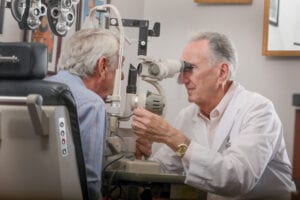
Our retina specialists, Dr. Leonard Kirsch, Dr. Richard Hairston, and Dr. Ho will determine the diagnosis and cause of cystoid macular edema by taking an extensive history and by performing a very detailed examination of your retina using special microscopes and lenses. The doctor may also recommend a test called a fluorescein angiogram. This test involves injecting a vegetable dye into a vein in the arm and taking black and white photographs of the retina using a special camera (these pictures are NOT X-rays). The photographs allow the doctor to confirm the diagnosis of cystoid macular edema, to determine where the leaky blood vessels are located, to discover how much leakage is occurring, and to guide treatment recommendations.
Treatment of Cystoid Macular Edema
 What will happen if we don’t treat clinically significant cystoid macular edema? If the edema fluid stays in the retina for many months, it can cause permanent damage to the macula and the vision may never be normal. Therefore, some form of treatment is recommended for almost everyone with significant cystoid macular edema.
The treatment that the specialists at The Eye Institute of West Florida recommend for cystoid macular edema is custom-designed for your individual situation. The treatment depends especially on the underlying cause of the cystoid macular edema. If you have cystoid macular edema following cataract surgery, the initial treatment (often started already by the cataract surgeon) is eye drops and perhaps an oral medication called Diamox. If the cystoid macular edema does not respond to this treatment, the next step involves an injection of prednisone medication behind the eye. This injection is done in the office after numbing the skin of the eye. The injections are usually painless, although the eye may feel swollen and sore for a day or two afterward. The eyelid may also become droopy but this will usually go away by itself. The doctor then allows 6 to 8 weeks for the medication to take effect and re-examines the retina.
Up to 3 injections may be required to cure the cystoid macular edema. Treatment with injections is successful in 80% to 90% of patients. If the cystoid macular edema is not cured using the injections behind the eye, injections of steroid medications and/or Avastin into the eye itself may be performed. Avastin is a medication that inhibits a molecule called vascular endothelial growth factor (VEGF) and is most commonly used for wet age-related macular degeneration. Recent clinical research has indicated that Avastin may be a safe and effective treatment for cystoid macular edema. Although it is used “off-label” for all eye diseases because it has only been approved to treat metastatic colon cancer, Avastin has become a standard of care for a number of eye problems over the past few years.
Any of these intraocular injections are also painless and performed in the office, although there is slightly more risk than incurred with the injections behind the eye. Risks of any injection into the eye include infection, bleeding, retinal detachment, cataract formation, and increased eye pressure (glaucoma); these are rare occurrences and can usually be treated. You will be given antibiotic drops for four days following this injection. The injections may be repeated about every 6 weeks if the edema persists or recurs. If the injections do not resolve the problem then surgery may be recommended.
 The operation for cystoid macular edema is performed in the regular operating room at the Largo Ambulatory Surgery Center and is almost always performed under local anesthesia. You may go home the same day or stay overnight in the hospital. The risks of this operation include retinal detachment (occasionally), infection inside the eye (rarely) and bleeding inside the eye (rarely). These complications are treatable most of the time, although as with any surgery inside the eye, loss of eyesight can rarely result.
The surgery is called a vitrectomy because the vitreous gel inside the eye is removed through 3 tiny incisions that do not require stitches. The vitreous gel is replaced with salt water (the vitreous is like your appendix; you have it but you are sometimes better off without it!). If there is scar tissue on the surface of the retina, the doctor will remove it during surgery. Using special microscopic forceps, the scar tissue is peeled from the surface of the retina like you would peel a label from its backing. You will wear an eye patch for the first night and must use drops for several weeks afterward. The operation has better than a 90% success rate.
Laser Treatment of Cystoid Macular Edema
For cystoid macular edema due to diabetes or a stroke in the retina, laser treatment is usually recommended as the first choice. Using a contact lens placed on the front of the eye and a state-of-the-art laser, the doctor places mild laser spots in the macula. The procedure is painless, takes less than 5 minutes, and is performed in the office. The major side effect is that some patients actually see the laser spots after the treatment is finished; these usually fade away over the next few weeks. The macular edema may take up to four months to go away. If the swelling does not go away after that time or it comes back later, the laser treatment can be repeated.
Maintaining Vision
As you can see, the diagnosis and treatment of cystoid macular edema is complex and is best performed by a retina specialist. We hope that you more clearly understand the different choices available and which may be the best for your situation. Working together with your surgeon, you can choose a treatment plan with which you are most comfortable and which has the highest chance of restoring and maintaining your precious gift of sight.
Schedule your Retina Evaluation today
Call (727) 581-8706 to schedule your appointment
Meet Your Retina Care Specialists
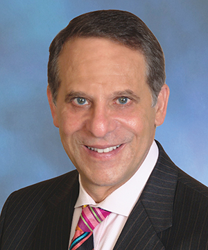
 Leonard S. Kirsch, M.D., F.R.C.S.(C) is a fellowship-trained vitreous and retina specialist. He is internationally known for the numerous papers and lectures he has presented here and abroad. Dr. Kirsch is currently an active participant in ongoing research to find new treatments for diseases of the retina. His expertise in the most advanced diagnostic and treatment techniques of all diseases of the retina, macula, and vitreous make Dr. Kirsch one of the elites in his field. In the Tampa Bay area, Dr. Kirsch pioneered the use of Photodynamic Therapy with Visudyne©, and intravitreal Macugen©, Lucentis©, and Avastin© for the treatment of age-related macular degeneration. Dr. Kirsch was also among the first surgeons in Florida to perform 25 gauge “no-stitch” vitrectomy in 2001. He is certified by the American Board of Ophthalmology and is a fellow of the Royal College of Surgeons of Canada.
 Richard J. Hairston, M.D. is a vitreous and retina specialist. He joined The Eye Institute in June 2001 coming to us from a busy retina practice in Washington, DC. Dr. Hairston graduated from the Johns Hopkins University School of Medicine and did his residency at the Wilmer Ophthalmological Institute at Johns Hopkins University. He completed a fellowship in diseases and surgery of the retina and vitreous at The Center for Retina Vitreous Surgery, Memphis, Tennessee. He then served as Assistant Chief of Service in Ophthalmology and Director of the Ocular Trauma Service at Johns Hopkins Hospital. Most recently he was Assistant Professor of Ophthalmology at Johns Hopkins University. He is certified by the American Board of Ophthalmology. Dr. Hairston enjoys an international reputation as an outstanding retina and vitreous surgeon.
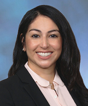
 Janie Ho M.D., is a board-certified ophthalmologist, fellowship-trained in medical and surgical vitreoretinal diseases such as macular degeneration, diabetic retinopathy and retinal tears and detachments. She has been educated at some of America’s finest institutions. She received her Bachelor of Arts from Harvard University and her medical degree from Duke University. She went on to ophthalmology residency at the University of California, San Francisco. Following residency, Dr. Ho continued on to a fellowship in vitreoretinal diseases at the prestigious Massachusetts Eye and Ear Infirmary of Harvard Medical School. Dr. Ho has participated in angiogenesis research, investigating causes and treatment for common retinal disorders.
 Sejal Shah M.D., is a board-certified, fellowship-trained Retina Specialist. Dr. Shah received her medical degree from the University of South Florida. She began her postgraduate training with an internship in medicine at UCLA, followed by ophthalmology training at the University of South Florida. She went on to complete a fellowship at the prestigious Bascom Palmer Eye Institute, specializing in the diagnosis and treatment of retinal disorders. Dr. Shah also earned a B.S. degree with honors in Nutritional Science from the University of Florida. Dr. Shah brings her expertise as a medical retinal specialist with skills in managing and treating vitreoretinal pathology which including age-related macular degeneration and diabetic retinopathy. Her research has appeared in publications such as Ophthalmic Surgery, Lasers, and Imaging Retina; Clinical Ocular Oncology; Survey of Ophthalmology; International Ophthalmology Clinics; and Survey of Ophthalmology. In her spare time, she enjoys spending quality time with her family and friends, traveling, and trying to keep up with her twin boys.

